



A plant gall is a remarkably well-structured habitat that the plant builds in response to another organism that alters its development at a specific site. This page explains how a puncture, an egg, or a tiny gall-inducing organism can trigger the formation of new plant tissue.
The key point in one sentence
What is a plant gall?
A plant gall is a localised area of newly formed or modified plant tissue. It develops in response to a gall inducer, such as an insect, mite, nematode, fungus, or bacterium. Galls can occur on leaves, buds, shoots, roots, or flowers. Some are barely visible; others form conspicuous spheres, spurs, or felt-like patches.
The precise form is often closely associated with the gall-inducing species and its host plant. A gall is more than an accidental injury: it is a plant developmental programme activated in an unusual place or at an unusual time.
What can induce galls?
Well-known gall inducers include gall wasps, gall midges, gall mites, aphids, and other sap-feeding insects. Caterpillars, sawflies, nematodes, fungi, bacteria, and some viruses can also cause gall-like growths. Not every swelling is a gall: injuries, diseases, and normal bud development can look similar.
The explanations below focus mainly on animal gall inducers; plant responses to microorganisms can follow different processes.
From first stimulus to mature gall
- A suitable site
The inducer selects a suitable plant species and young, responsive tissue. - Egg-laying or feeding
A puncture, egg-laying, saliva, and repeated feeding stimuli act together at the site. - Signals reach the cells
Mechanical stimuli and released compounds alter plant cell responses. - Growth is redirected
Hormone and defence signalling and gene activity shift, prompting cells to divide or enlarge. - The gall develops
The plant forms protective and nutritive tissue. A larva or mite often lives and feeds inside.
This is a simplified model. Depending on the inducer, plant, and gall type, the steps may vary in intensity or timing.

What does the gall inducer do?
Mechanical stimuli
Wasp oviposition, punctures, rasping or sucking during feeding, and larval movements can injure plant cells and trigger pressure or wound signals. Repeated contact and feeding at a small site may also matter. Mechanical stimulation alone cannot explain the precise form of many galls.
Secretions and other chemical signals
While feeding or laying eggs, gall inducers release saliva, secretions, or excretions. These may contain compounds that alter plant responses. Some systems provide evidence for hormones or hormone-like substances, but the key molecules remain unknown for many gall wasps, gall midges, and gall mites. No single compound should be assumed to explain all galls.
An interaction, not a single trigger: A gall usually arises through interactions among the host plant, gall inducer, tissue age, location, and timing. Signals from the inducer influence plant regulation; living plant cells build the gall.
Hormones and plant signalling pathways
Plant hormones are signalling molecules that regulate growth, development, and defence. No single “gall hormone” is responsible for all galls. Local concentrations, transport, and interactions among multiple signals are key.
Auxin
Regulates cell elongation, cell division, and vascular tissue arrangement, among other processes. Altered auxin production or distribution has been observed or proposed in many gall types.
Cytokinin
Often promotes cell division and can affect how long cells remain capable of dividing. It acts in close interaction with auxin.
Jasmonate & salicylic acid
Both are important plant defence signals. Depending on the system, their effects in galls may be weakened, redirected, or locally restricted.
Ethylene
A gaseous signalling molecule involved in stress responses, ripening, and growth. Its role depends greatly on the plant and gall inducer.
ROS
Reactive oxygen species (ROS) are short-lived, highly reactive molecules. At controlled concentrations, they also act as signals in wound, defence, and developmental responses.
Gene activity
Signals can switch genes on or off, changing which proteins and structural components a cell produces and what role it performs.
How does plant tissue change?
- More cell division – hyperplasia: Additional cells form, for example in growing gall layers.
- Larger cells – hypertrophy: Individual cells swell or grow beyond their normal size.
- New tissue organisation: Cells differentiate into distinct tissue types; the gall may develop an outer layer, nutritive tissue, and a larval chamber.
- Cell walls and tissue firmness: Cell walls may thicken, strengthen, or undergo chemical changes, affecting protection, shape, and firmness.
- Vascular tissue and nutrient supply: In many galls, vascular bundles change or new ones form. Water, sugars, and other nutrients are directed to the gall.
- Metabolism: Sugars, amino acids, and other compounds may accumulate locally. Pigments, tannins, and defence compounds also change.
Why is a gall a nutrient sink?
A nutrient sink is tissue that attracts and consumes resources rather than simply transporting them onward. The plant may supply the gall with sugars and other nutrients. This provides an accessible food source for the larva and may also offer a protected chamber and favourable microclimate. The gall generally provides the inducer with food, protection, and a place to develop.
The plant does not automatically benefit. Galls can impose a cost or deform leaves and shoots. Some cause little harm; the effects of others depend on their number, the plant species, and environmental conditions.
Gall mites: tiny inducers, striking forms
Gall mites are microscopic, worm-shaped arachnids. Many are associated with particular plant species or even specific plant parts. As they feed, they release compounds and expose sensitive young cells to localised stimuli. Leaves may respond by developing felt-like patches, small pouches, or spurs. Some gall mites live in buds and cause enlarged, rosette-like structures.
In erineum galls, unusually large numbers of plant hairs (trichomes) grow on the leaf underside. These dense hairs form a felt-like covering and provide mites with a sheltered habitat. Nail galls on lime leaves, by contrast, are small, pointed projections from the leaf surface. These are distinct forms, and not every visible gall is produced by the same mite species.
Gall forms – examples from nature
The drawings are botanical illustrations created specifically for this page. They show typical examples schematically; size and colour may vary in nature.
| Gall form | Illustration and typical example | What does it look like? | Example gall inducer |
|---|---|---|---|
| Erineum gall | 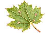
|
Dense plant hairs form small felt-like patches, often on the leaf underside. | Gall mites, e.g. erineum gall mites on maple (Acer). |
| Pouch gall | 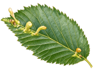
|
Leaf tissue bulges upwards or downwards into a pouch-like shape. | Aphids, e.g. the elm-cockscomb gall aphid Tetraneura ulmi. |
| Rolled leaf margin | 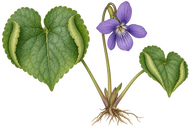
|
The leaf margin rolls inwards and may become thickened or fleshy. | Gall midge Dasineura odoratae on sweet violet (Viola odorata). |
| Nail gall | 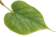
|
Short, pointed, often reddish or yellowish projections on the upper leaf surface. | Gall mites on lime (Tilia), such as Eriophyes tiliae. |
| Spherical gall (oak apple) | 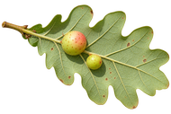
|
A rounded, fleshy gall that may later become firm, often with an internal chamber. | Gall wasps (Cynipidae) on oak (Quercus). |
| Bud gall | 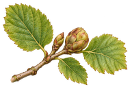
|
A bud enlarges, remains closed, or develops a rosette-like form. | Hazel bud gall mite Phytoptus avellanae. |
| Shoot gall | 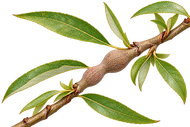
|
A twig or stem thickens into a knot-like or spindle-shaped swelling. | Leaf-mining fly Hexomyza simplicoides on willow (Salix). |
| Encircling gall | 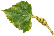
|
The petiole grows spirally around and encloses an aphid colony. | Poplar petiole gall aphid Pemphigus spyrothecae on poplar (Populus). |
What do we know, and what remains uncertain?
Well established
Galls consist of plant tissue. Many show increased cell division, enlarged cells, altered gene activity, and targeted nutrient supply. Plant hormones, particularly auxin and cytokinin, and defence signals are involved in numerous systems. Gall inducers often benefit from food and protection.
Ongoing research
For many species, the initial triggering molecules remain unknown. It is not always clear whether hormones come from the inducer, are produced by the plant, or involve both. How a gall wasp, gall midge, or gall mite directs the characteristic form remains unresolved for many galls. There is probably no single universal mechanism.
References and further information
- Harris, M. O. & Pitzschke, A. (2020): Plants make galls to accommodate foreigners: some are friends, most are foes. New Phytologist. doi:10.1111/nph.16340.
- Huang, M.-Y. et al. (2021): Recent Progress Regarding the Molecular Aspects of Insect Gall Formation. International Journal of Molecular Sciences 22:942. Full text (PMC).
- Suzuki, Y. & Tokuda, M. (2023): Insect gall induction and phytohormones. Regulation of Plant Growth & Development 58:105–111. doi:10.18978/jscrp.58.2_105.
- Tooker, J. F. & Helms, A. M. (2014): Phytohormone dynamics associated with gall insects, and their potential role in the evolution of the gall-inducing habit. Journal of Chemical Ecology 40:742–753. doi:10.1007/s10886-014-0457-6.
- Royal Horticultural Society: Lime nail gall mite – Nail galls on lime leaves. RHS.
- Bavarian State Institute of Forestry: information on erineum galls on maple and Aceria cephalonea on field maple. LWF Bayern; Field maple article (PDF).
- INRAE Ephytia: Phytoptus avellanae, the hazel bud gall mite. INRAE.
- Dasineura odoratae on Viola odorata: “Gales de Catalunya” species account. Species account.
The illustrations are original schematic drawings, not photographs or images to scale. Scientific information current to October 2026.